
Free Printable Human Eye Diagram worksheets help kids explore how the eye works through labeling activities, coloring pages, quizzes, and hands-on crafts. Download and print PDF diagrams, vocabulary mats, and research pages covering everything from simple eye parts for young learners to detailed vision pathway and light diagrams for older students. These worksheets build skills in anatomy vocabulary, diagram reading, and scientific reasoning while keeping the content playful and easy to follow. Teachers can use them for science centers or whole-class lessons, and parents will find them just as useful for homeschool units on the human body.
Simple Human Eye Diagrams for Kids
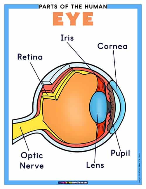
Download and print this simple eye diagram for kids. This colorful chart labels six basic interior parts of the human eye, giving young learners an easy introduction to eye anatomy.
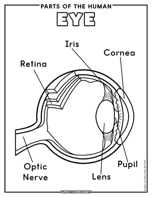
Simple Eye Diagram B&W Coloring Page
This labeled eye diagram is a black-and-white version of our simplified chart above. Covering six basic interior parts, it makes a great coloring activity for early learners.
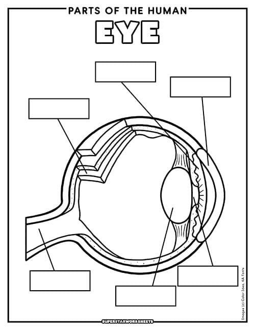
Download and print this simple blank labeling diagram of the human eye. Students write the name of each part in the provided blank box, reinforcing basic eye vocabulary.
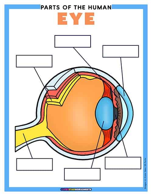
This learning center comes with printable prewritten labels for students to place on the printable mat. This low-risk activity provides ample practice for locating and naming the basic parts of the eye.
Printable Human Eye Diagrams
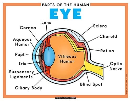
Download and print this free Parts of the Human Eye Diagram for your students. Featuring a large, color-coded anatomical illustration of the eye with 13 labeled parts. For more FREE human science worksheets, check out our Heart Worksheets today!
Human Eye Diagram Black & White
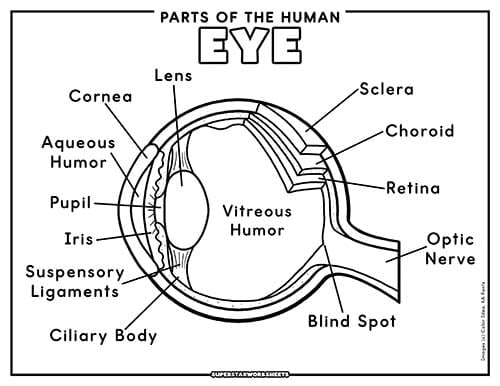
Free printable human eye diagram with a simple outline illustration. This PDF is labeled with the parts of the eye and is great for students to use as a cross-section reference.
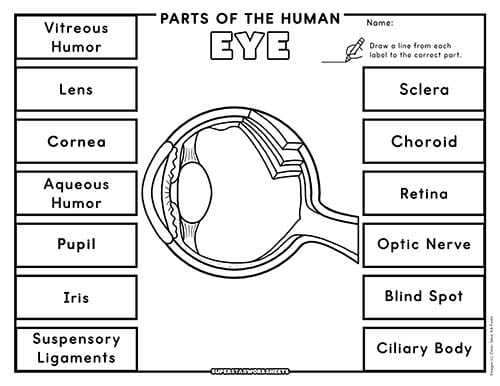
Students can begin to learn the part names and locations with this printable human eye labeling worksheet. Students draw a line from each label to the correct part on the diagram. Teachers! Check out these FREE Ear Worksheets today!
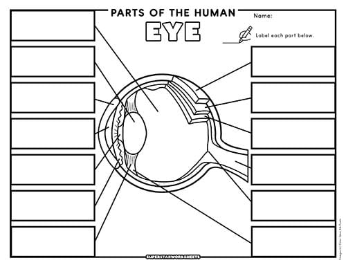
Blank Human Eye Labeling Diagram
Students must recall the names of each of the 13 main parts of the human eye and write them on the blank lines provided. This is a good way to assess student retention.
Eye Anatomy Quizzes
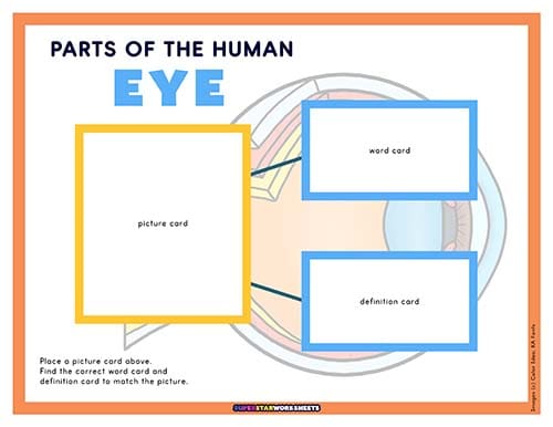
Our take on traditional Montessori Nomenclature cards will optimize your learning centers with simple visual illustrations, bright highlights on individual parts, and self-checking picture, word, and definition cards.
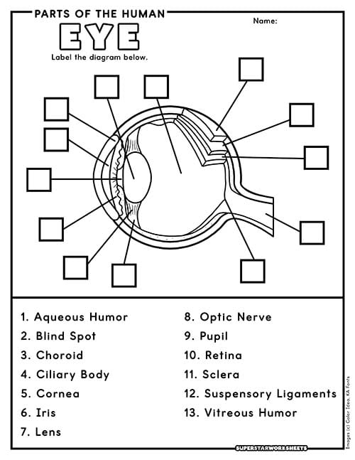
Assess student knowledge with this free printable human eye labeling quiz. Featuring 13 numbered parts, students write the correct term on the line, with an answer key included for easy grading. Teachers! Take a look at our FREE Lungs Worksheets!
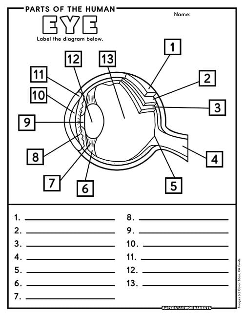
Assess student knowledge with this free printable human eye labeling quiz. Featuring 13 numbered parts, students write the correct term on the line, with an answer key included for easy grading.
Human Eye Anatomy Printables
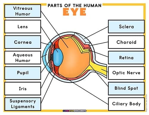
Here is a full-color labeled human eye diagram that makes a great reference chart or poster for your science classroom.
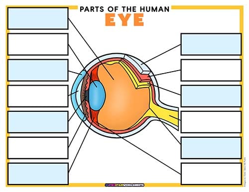
Download this printable learning center activity mat for students who are learning the location and function of the parts of the human eye. This set includes printable label cards as well as definition cards for an extra challenge.
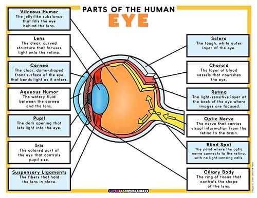
Here is a full-color labeled human eye diagram that traces how blood reaches the eye through the carotid and ophthalmic arteries, making a great reference chart for your science classroom.
Printable Eye Vision Diagrams
Aqueous Humor: The clear, watery fluid that fills the space between the cornea and the lens.
Blind Spot: The point where the optic nerve connects to the retina, where there are no light-sensing cells.
Choroid: The layer of blood vessels between the retina and the sclera that supplies the eye with oxygen and nutrients.
Ciliary Body: The ring of tissue that contains the ciliary muscle and controls the shape of the lens.
Cornea: The clear, dome-shaped front surface of the eye that bends light as it enters.
Iris: The colored part of the eye that controls how much light enters by adjusting the size of the pupil.
Lens: The clear, curved structure behind the pupil that focuses light onto the retina.
Optic Nerve: The nerve that carries visual information from the retina to the brain.
Pupil: The dark, round opening in the center of the iris that lets light into the eye.
Retina: The light-sensitive layer at the back of the eye where images are focused, containing the rods and cones.
Sclera: The tough, white outer layer that protects and gives shape to the eye.
Suspensory Ligaments: The tiny fibers that hold the lens in place and help it change shape.
Vitreous Humor: The clear, jelly-like substance that fills the large space behind the lens and helps the eye hold its round shape.
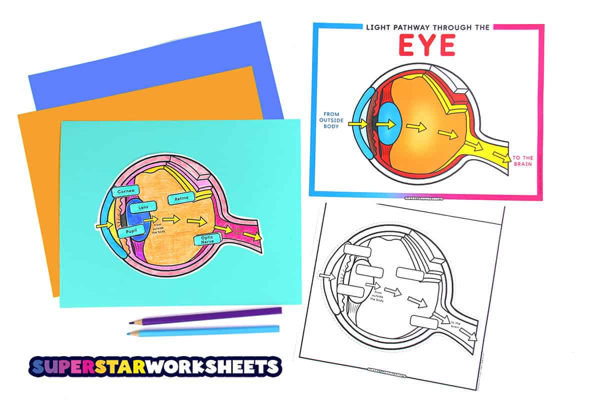
Printable Eye Vision Diagrams
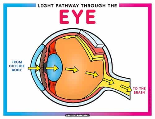
This large diagram traces the pathway of light through the eye using a color-coded arrow system, from outside the body all the way to the brain. It is an ideal anchor chart for teaching how vision works.
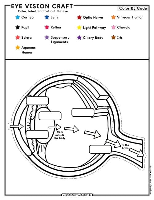
Eye Vision Color by Code Craft
Students color and assemble this hands on craft using a color code that matches the light pathway diagram. It turns the process of seeing into a memorable, take home project.
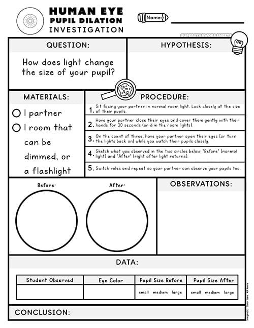
Human Eye Pupil Dilation Investigation
This simple partner experiment lets kids observe how their pupils change size in response to light. It is a quick, low-mess way to see the eye’s light response in action.
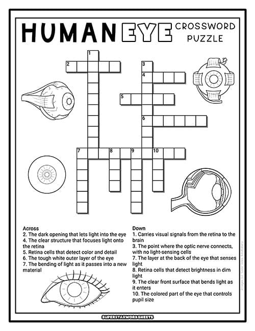
This crossword puzzle reinforces eye vocabulary using clues pulled from the anatomy and vision vocabulary banks. It makes a great review activity after an eye unit.
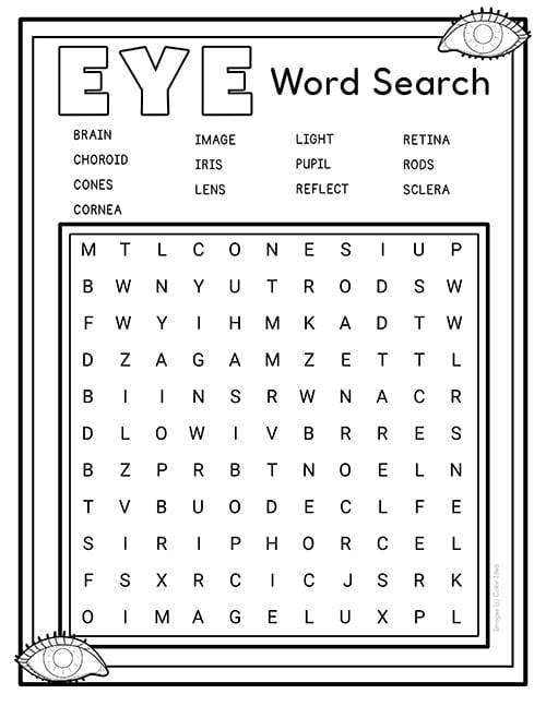
This word search gives students a fun, low-pressure way to recognize key eye and vision vocabulary. It works well as an early finisher or homework activity.
How the Eye Works
The eye has one main job: turn light into a picture your brain can understand. Light bounces off everything around you and enters the eye through the clear cornea, then passes through the pupil and lens, which bend and focus it onto the retina at the back of the eye. Special cells called rods and cones sense that light and turn it into signals the optic nerve carries straight to the brain. The picture that lands on the retina is actually upside down, so the brain flips it right side up before you ever notice a thing. Working together, the eye and brain complete this entire process in a fraction of a second, nearly every moment you are awake.
Human Eye Facts for Kids
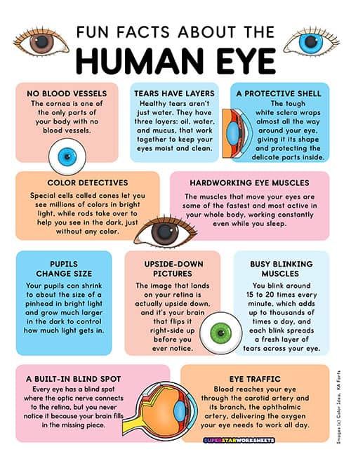
Fun Facts About the Human Eye for Kids
Download and print this fun facts poster with amazing information about the human eye. These are great charts to keep near your science learning centers for quick reference.
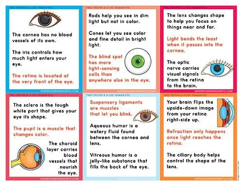
Here is a fun printable game you can play with your students. Two Truths and a Lie is a fact-finding game. Each card contains two truths about the human eye and one lie for students to spot.
This printable graphic organizer will help students keep track of their human eye research. Students fill in each section of the KWL+Q research guide.
Human Eye: Fun Fact Finder (Research)
Encourage your students to seek and find fun facts about the human eye with this printable research-based scavenger hunt. Students will put their research skills to work across nine open-ended prompts.
Fun Facts About the Human Eye
No Blood Vessels: The cornea is the only part of your body with no blood vessels at all. It gets the oxygen and nutrients it needs straight from your tears and the fluid behind it.
A Built-In Blind Spot: Every eye has a blind spot where the optic nerve connects to the retina, but you never notice it because your brain fills in the missing piece.
Pupils Change Size: Your pupils can shrink to about the size of a pinhead in bright light and grow much larger in the dark to control how much light gets in.
Upside-Down Pictures: The image that lands on your retina is actually upside down, and it’s your brain that flips it right-side up before you ever notice.
Busy Blinking Muscles: You blink around 15 to 20 times every minute, which adds up to thousands of times a day, and each blink spreads a fresh layer of tears across your eye.
Hardworking Eye Muscles: The muscles that move your eyes are some of the fastest and most active in your whole body, working constantly even while you sleep.
Color Detectives: Special cells called cones let you see millions of colors in bright light, while rods take over to help you see in the dark, just without any color.
Tears Have Layers: Healthy tears aren’t just water. They have three layers- oil, water, and mucus- that work together to keep your eyes moist and clean.
A Protective Shell: The tough white sclera wraps almost all the way around your eye, giving it its shape and protecting the delicate parts inside.
Two-Way Traffic: Blood reaches your eye through the carotid artery and its branch, the ophthalmic artery, delivering the oxygen your eye needs to work all day.
Leave a Reply